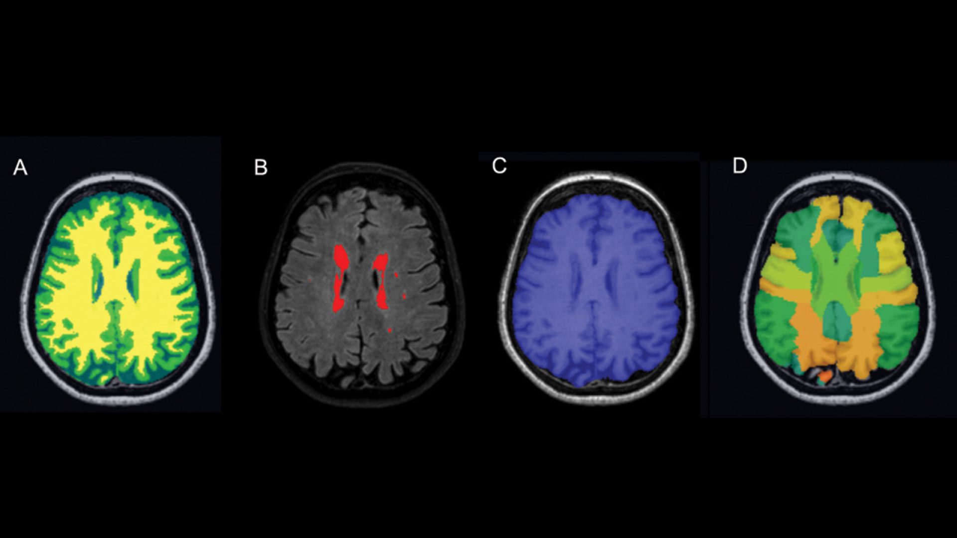
Fig. 1. Imaging biomarkers of the structural brain scan of the Heart-Brain Study: A) brain tissue segmentation (yellow = white matter (WM), green = gray matter (GM), blue = cerebrospinal fluid (CSF), B) red = white matter hyperintensities (WMH), C) brain mask, D) 83 regions-of-interest.
Fig. 2. Arterial spin labeling (ASL) scans for one Heart-Brain Study participant, four axial slices are shown. A) Perfusion-weighted ASL images after motion correction, B) cerebral blood flow (CBF) map, C) CBF map as color-overlay with M0 normalization image as background.
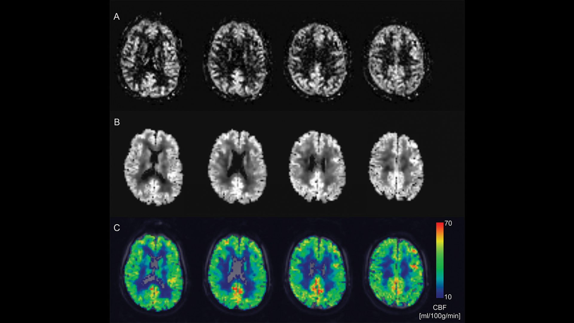
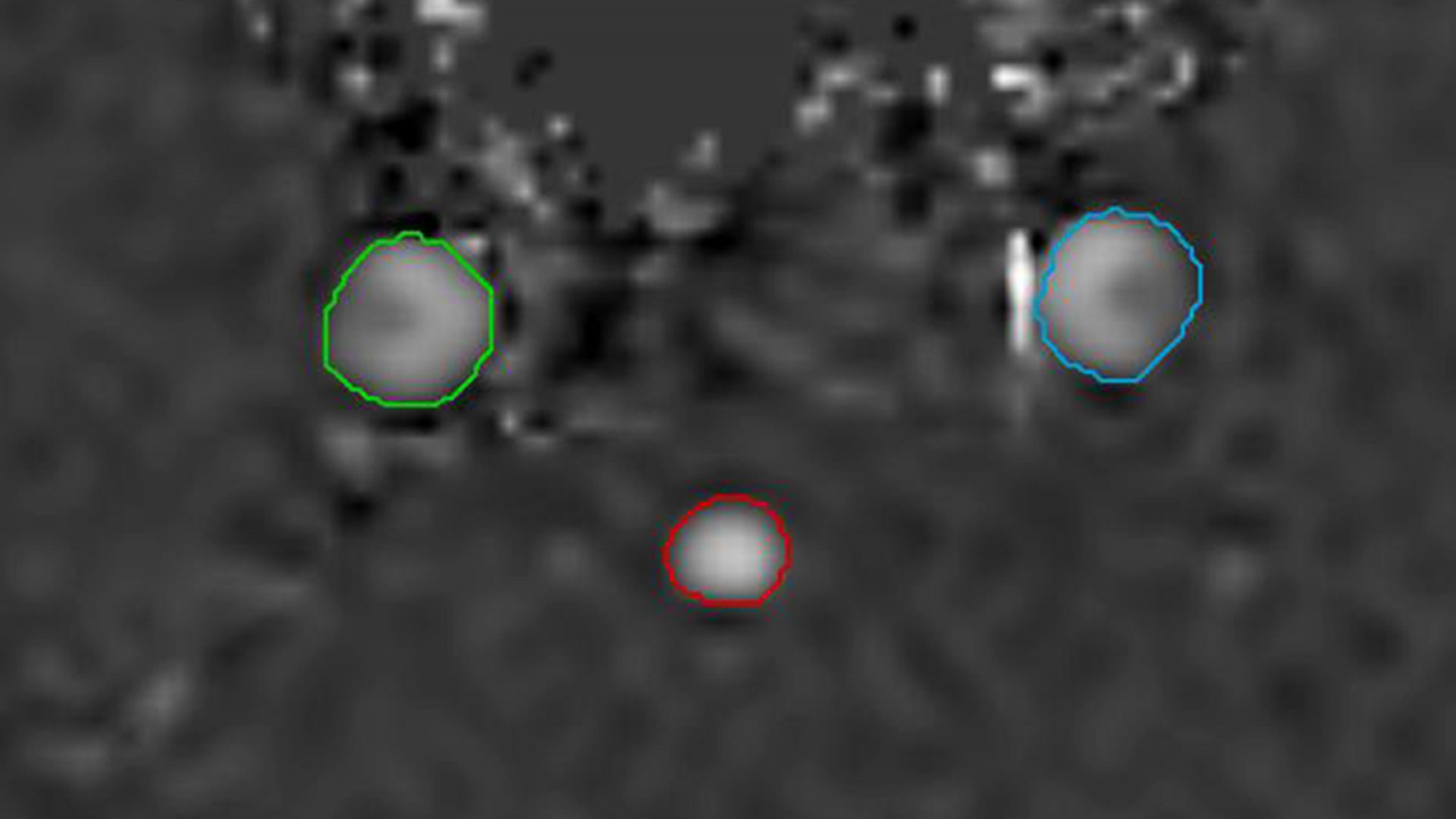
Fig. 3. Q-flow pipeline; manual segmentation of left and right carotid artery and basilar artery.
Fig. 4. Examples of MR cardiac parameters that are extracted through automated cardiac post-processing. An automatic pipeline for epicardium and endocardium contour detection on the two chamber (below) and four chamber (above) cardiac MR cine scans. The proposed pipeline uses multi-atlas-based segmentation and an image registration based model for the contour detection. These contours are used to process short-axis scans for true volumetric parameters.
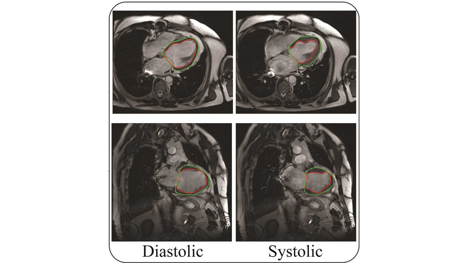
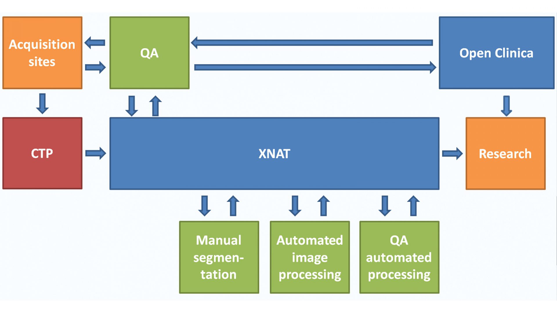
Fig. 5. Overview of the data storage and analysis platform and data flow. MRI scans are uploaded from the four data acquisition sites to Extensible Neuroimaging Archive Toolkit (XNAT). For anonymization of the scans, data is sent via a Clinical Trial Processor (CTP). The consistency of the data in XNAT and their correspondence with the clinical data in OpenClinica is assessed weekly with an automated quality assessment (QA) procedure. In addition, scans are downloaded from XNAT for manual and automatic processing, and for visual QA of the scans and the automated processing results. Results of all steps are uploaded back to XNAT. Finally, derived image data are downloaded from XNAT and clinical data are downloaded from OpenClinica. For analysis, downloaded data are combined into appropriate software formats.
Images from:
Hooghiemstra AM, Bertens AS, Leeuwis AE: et al. The Missing Link in the Pathophysiology of Vascular Cognitive Impairment: Design of the Heart-Brain Study. Cerebrovasc Dis Extra 2017;7:140-152. doi: 10.1159/000480738
https://pubmed.ncbi.nlm.nih.gov/29017156/